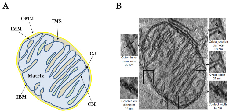
Figure 1.
Structure of mitochondrial microcompartments. (A) Schematic representation of mitochondrial architecture. The outer mitochondrial membrane (OMM), inner mitochondrial membrane (IMM), inner boundary membrane (IBM), cristae junctions (CJ), intermembrane space (IMS), cristae membrane (CM) and mitochondrial matrix are indicated. (B) Electron micrograph from a cryo cut mitochondrion with antibody probing of OXPHOS complexes. The localization in the cristae membrane is obvious. Inset: detailed view of the two IMM compartments CM and IBM connected by the cristae junction (CJ). Scale bar: 150 nm. Reproduced with permission from Frey et al. [15], Trends Biochem. Sci.; published by Elsevier, 2000.

