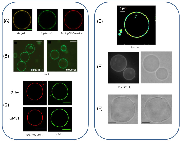
Figure 10.
Giant vesicles as model systems. (A) Confocal image of a GUV made of EPC/CL 60:40 mol% showing the distribution of Top-Fluor CL (green channel) and Bodipy-TR Ceramide (red channel), 22 C, scale bar is 5 µm, [80]. (B) Confocal microscopy of GUVs made of PC/CL 90:10 and 80:20 mol%, room temperature, scale bar is 50 µM, [139]. (C) GUVs made of composed of (18:0–22:6)PC/(16:0–20:4)PE/(18:2)CL/DOPI/DOPS/Chol (39.9:30:20:5:3:2 mol% and giant mitochondrial vesicles (GMVs) from native phospholipids extracted visualized by Texas Red DHPE (left) and by NAO (right) at 0.1 mol%. room temperature, scale bars are 10 µm, [100]. (D) Laurdan GP image of isolated EPC/EPE/CL (50:25:25) mol% membrane, room temperature, scale bar is 5 µm, [141]. (E) Fluorescence and optic microscopy images of POPC/DOPE/CL (49:30:20) GUV containing 0.5 mol% TopFluor-CL, 25 C, [140]. (F) Phase contrast images of GUVs made of POPC/CL (70:30) mol%, 23 C, [142]. Reproduced with permissions from respectively Beltrán-Heredia et al., Pennington et al., Jalmar et al., Kawai et al., Cheniour et al., and Tomšié et al. [80,100,139,140,141,142], in respectively Commun. Biol., J. Biol. Chem., Cell Death Dis., J. Phys. Chem. B, Biochim. Biophys. Acta, and J. Chem. Inf. Model; published respectively by Nature Publishing Group (2019), Elsevier (2018), Nature Publishing Group (2010), ACS (2014), Elsevier (2017), and ACS (2005).

