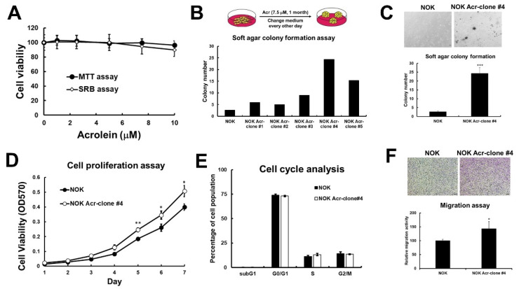
Figure 1.
Acrolein induced cell transformation in normal human keratinocytes (NOK). (A) Cell viability of NOK under low dose of acrolein (0–10 μM) treatment for 1–3 days was analyzed using MTT assays and SRB assays. (B) NOK cells were treated acrolein (Acr, 7.5 μM) for one month and named as Acr-clone #1–5. Anchorage independent cell growth of NOK Acr-clone #1–5 was analyzed using soft agar assays. (C) Soft agar anchorage-dependent cell growth of NOK Acr-clone #4 was analyzed using soft agar assays. (D) Cell proliferation of NOK Acr-clone #4 was analyzed using MTT assays. (E) Cell cycle progression of NOK Acr-clone #4 was analyzed using cell cycle analysis with PI staining. (F) Cell migration activity of NOK Acr-clone #4 was analyzed using transwell migration analysis. Data were presented as the mean ± s.d. Student’s t-tests were used to determine statistical significance, and two-tailed p-values are shown. *** p < 0.005, ** p < 0.01, * p < 0.05 compared with NOK parental cells.

