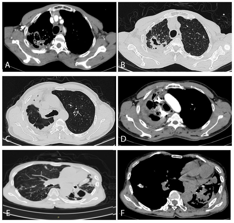
Figure 3.
Typical computed tomography findings in our CPA patients. (A,B) were from a 71-year-old male patient, (C,D) were of a 50-year-old male patient, and (E,F) were from a 78-year-old female patient. (A) (contrast enhanced scan) shows enlarged arterial vessels on the edge of two separate cavities posteriorly in the right lung. The lung windows from a slightly higher section show a large thick-walled cavity with an irregular interior lining, and probably three other much smaller cavities, in association with remarkable pleural thickening posteriorly, with some pleural fat latero-posteriorly. No aspergilloma is visible in either image. (C) shows extensive pleuro-pulmonary fibrosis encasing the right upper lung, with two small cavities (probably) anteriorly. The right main bronchus and mediastinum is shifted to the right. In (D), slightly higher in the chest at the level of the aortic arch, shows considerable major arterial blood vessel distortion, additional enlarged arteries within the areas of inflammation or fibrosis and an anterior cavity. (E) shows at least one thick-walled cavity at the top of the left overlying an area of significant pleural thickening, with areas of consolidation or fibrosis anteriorly, containing some calcification on a bullous emphysematous background. There is a small area of ill-defined inflammation in the right lung. The bronchial walls contain significant calcification. (F) also shows the major mediastinal shift to the left, with extensive areas of consolidation or fibrosis with no particular pattern.

