Fig. 4.
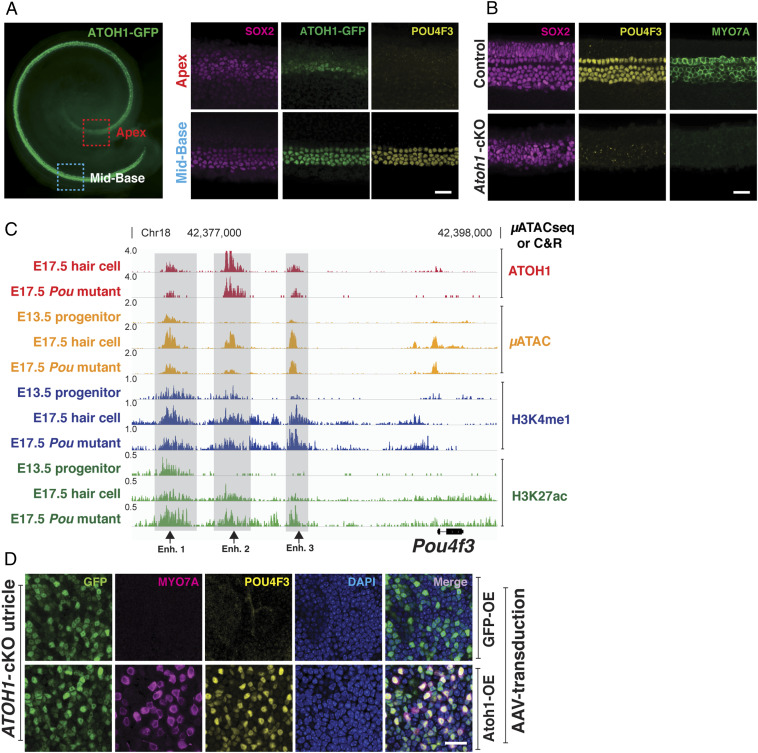
ATOH1 and POU4F3 synergize to form a feed-forward regulatory circuit in differentiating hair cells. (A) Whole mount of developing cochlea expressing ATOH1-GFP (green) at E15.5 (Leftl; red dashed rectangle, apical region; blue dashed rectangle, midbasal region). (Scale bar, 100 μm.) Higher-magnification images indicate that in the midbase, where hair cell differentiation first initiates, ATOH1 and POU4F3 are coexpressed; while in the apex, ATOH1 is expressed, but POU4F3 expression is not detected at this time within the basal-apical gradient characteristic of cochlear development. (Scale bar, 20 μm.) (B) Midbasal region of the cochlea at E15.5 in both wild-type and Atoh1 conditional knockout (Pax2-Cre) is shown. No hair cells are formed in the Atoh1 conditional knockout cochlea (MYO7A, green), and POU4F3 (yellow) is not expressed. (Scale bar, 20 μm.) (C) Genome browser (IGV) representation of the μATACseq and C&R results at the Pou4f3 locus. Putative ATOH1-bound enhancers for Pou4f3 up-regulation in the hair cells are indicated (gray bars). ATOH1-bound enhancers 1 and 3 are accessible in progenitors and labeled by H3K4me1 (black arrows). Binding of ATOH1 to all three indicated sites is not affected in Pou4f3 mutant. These data suggest that Pou4f3 expression in the hair cells is directly controlled by ATOH1. (D) Confocal images of utricular explant from Atoh1 conditional knockout mice. The utricles are overexpressed with GFP (control) or ATOH1-GFP, encoded in the AAV. Overexpression of ATOH1 but not GFP (control) induced the expression of POU4F3, suggesting that ATOH1 is sufficient for Pou4f3 expression in the inner ear. (Scale bar, 20 μm.)

