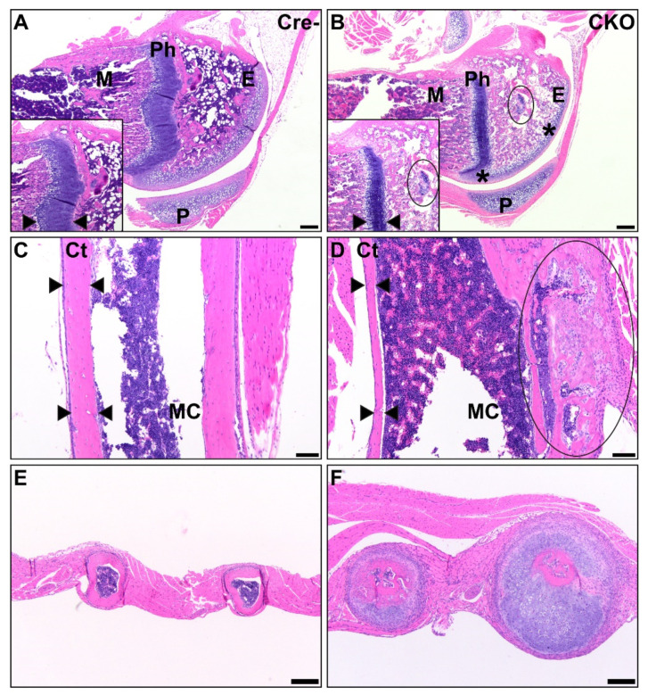
Figure 4.
CKO mice exhibit osteochondral dysplasia in long bones (B,D,F) compared to Cre- age-matched controls (A,C,E). A/B: In the femur, there was thin and sclerotic articular cartilage (asterisk), sparse epiphyseal trabeculae with retained cartilage cores (circle), thin and disorganized physes, and decreased metaphyseal primary spongiosa present in CKO mice (B) compared to control mice (A). Insets show a comparison of the physis thickness, with margins demarcated by arrows, and the retained cartilage core. Scale bars = 200 µm. E: epiphysis, Ph: physis, M: metaphyseal primary spongiosa, P: patella. C/D: In the humeral diaphysis, CKO mice (D) had markedly thinner cortices compared to age-matched Cre- control mice (C). CKO mice had occasional subperiosteal growths (circle) of cartilage, woven bone, remodeling cortical bone, and precursor-like cells. The cortex thickness is delineated by arrows. Scale bars = 100 μm. Ct: cortex, MC: medullary cavity. E/F: The ribs of a CKO mouse (F) were many times larger than those of a comparable Cre- control mouse (E) due to excessive cartilage surrounding a central core of irregular bone. Scale bars = 200 μm.

