Figure 6.
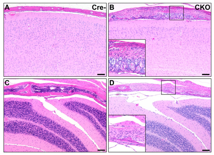
Osteochondral dysplasia in multiple bones of the calvaria in CKO mice. Compared to Cre- age-matched control mice (A,C), multiple bones were affected in CKO mice, including the parietal bone (B) and interparietal bone (D). Lesions similar to the osteochondral dysplasia observed elsewhere and consisting of variable amounts of bone, cartilage, fibrous connective tissue, and precursor-like cells were present in some areas (B), while in others, the bone was segmentally absent, with the brain encased only by fibrous connective tissue and precursor-like cells (D). Insets in (B,D) provide higher magnification views. The photos shown in (A,B) were taken mid-parietal at the level of the middle hippocampus, while (C,D) were taken mid-interparietal over the cerebellum. Scale bars = 100 μm.

