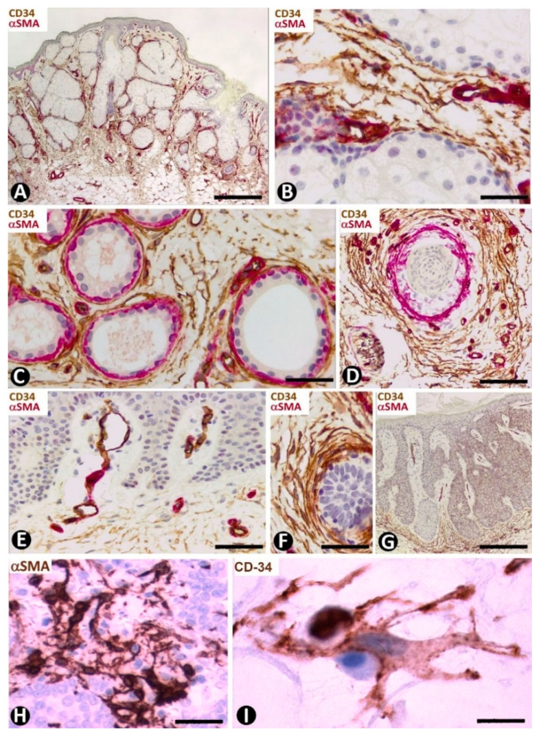
Figure 16.
Nevus sebaceous of Jadassohn (A–F), seborrheic keratosis (G) and Merkel cell carcinoma (H,I). (A–G) Double immunochemistry for CD34 (brown) and αSMA (red). Haematoxylin counterstain. (H,I) Immunochemistry for αSMA (H) and CD34 (I). (A) Panoramic view showing hyperplasia of superficially located sebaceous glands. Note that one gland opens through the epidermis. (B–D): Numerous CD34+SCs/TCs around sebaceous glands (B), sweat glands (C) and an involutive hair follicle in the lesion (D). (E) Absence of perivascular and interstitial CD34+SCs/TCs in areas with papillomatosis. (F) Abundant CD34+SCs/TCs around a basaloid proliferation. (G) Seborrheic keratosis in which CD34+ stromal cells are located at the bottom of the lesion, but are not observed in papillary areas with papillomatosis. (H,I) Merkel cell carcinoma with stromal cells, which show an increased somatic region and voluminous nuclei, and express αSMA (H) and CD34 (I). Bar: (A,G) 150 µm, (B,H) 45 µm, (C–F) 80 µm, I 15 µm.

