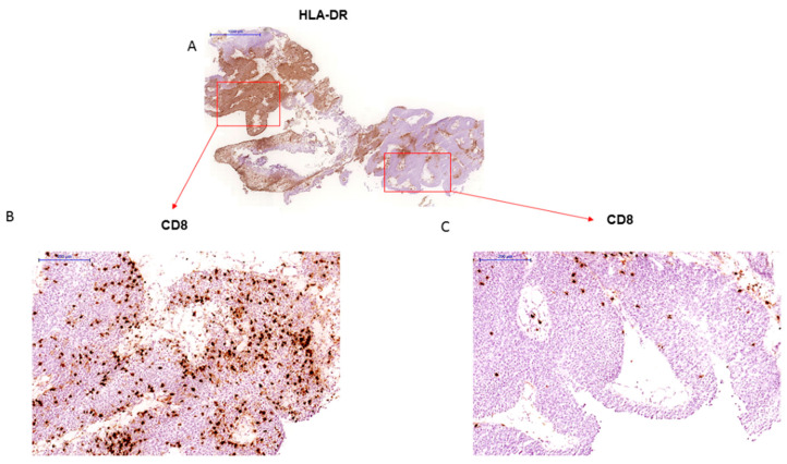
Figure 2.
(A) Bladder tumor sample with heterogeneous pattern of HLA-DR immunostaining (20× magnification) (B) HLA-DR positive area is heavily infiltrated with CD8+ T-cells (“inflamed” tumor), while HLA-DR negative distal zone is significantly less infiltrated with more lymphocytes in the stroma at the tumor margin (C) (A and C–at a 100× magnification).

