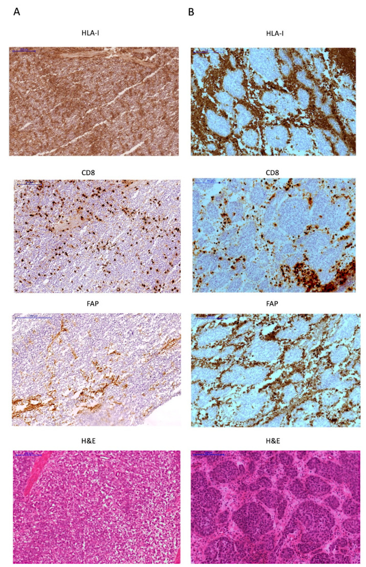
Figure 4.
Two different examples of FAP expression in the stroma in high grade tumors. (A) HLA-I/PD-L1 double positive tumor heavily infiltrated with T-cells, with few FAP+ stroma cells diffusely distributed in the tumor (B) HLA-I negative/PD-L1 positive “encapsulated” tumor with abundant FAP+ cells surrounding HLA-I negative tumor nest and CD8+ T-cells confined in the peritumoral stroma. All images are at 100× magnification.

