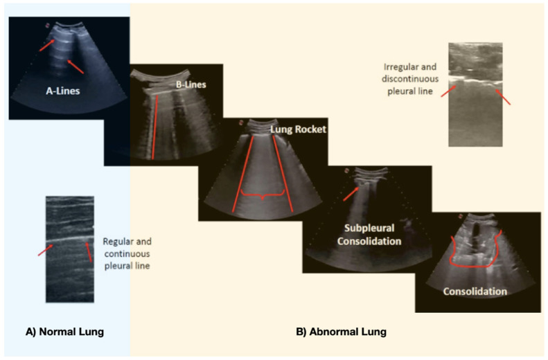
Figure 2.
Main lung ultrasound findings in COVID-19 pneumonia. (A) Normal lung pattern of horizontal (red arrow) lines parallel to pleura (A-lines). (B) Abnormal Lung: B-lines: Pattern of vertical lines that reach the depth of field and start from the pleural line (red line). Lung Rocket: A “white lung” (braces) where the B lines converge inside an intercostal space (rib shadow—red lines). The pleural line is usually fragmented and irregular. If the pleural line increases the irregularity, it might generate a subpleural consolidation. If the subpleural consolidation (<1 cm) progresses, or in superinfection cases, big consolidations (>1 cm) appear.

