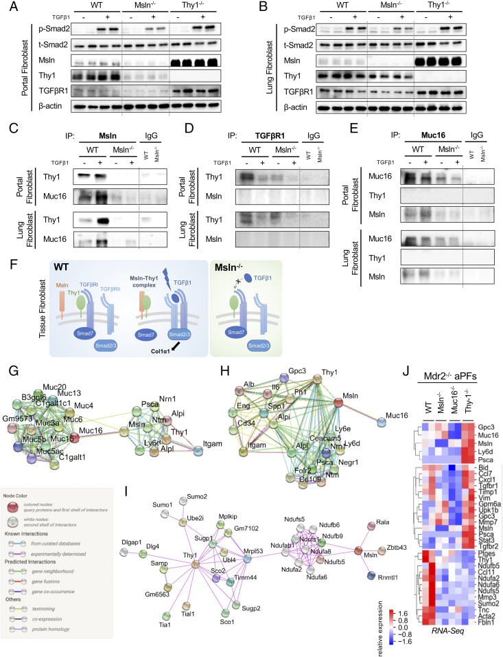
Fig. 5.
Msln regulates similar responses in aPFs and lung fibroblasts. (A–E) Responses to ±TGFβ1 (5 ng/mL, for 30 min) were compared in aPFs (Mdr2−/−, Msln−/−Mdr2−/−, and Thy1−/−Mdr2−/−) (A) and activated lung fibroblasts (WT, Msln−/−, and Thy1−/−) (B) using Western blotting for phospho-Smad2/Smad2, Msln, Thy1, and TGFβRI (normalized to the levels of β-actin expression), or IPs with anti-Msln Ab (C), anti-TGFβRI Ab (D), and anti-Muc16 Ab (vs. IgG) (E). (F) Proposed model of Msln–Thy1–TGFβRI signaling in tissue fibroblasts (see explanations in the text). (G–J) STRING network analysis depicts the top 20 first-neighbor genes connecting Msln–Thy1 (G) and Muc16–Msln (H). Known (shown with turquoise and purple connecting lines), predicted (green, red, and blue lines), or other (lime, black, and dark blue lines) interactions are shown. (I) Top 30 first- and second-neighbor genes connecting Msln–Thy1, experimentally determined interactions only. (J) RNA-seq–based relative expression of selected genes in Mdr2−/−, Msln−/−Mdr2−/−, Muc16−/−Mdr2−/−, and Thy1−/−Mdr2−/− aPFs is shown.

