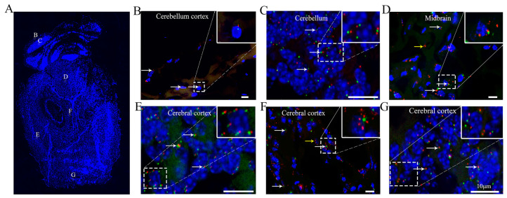
Figure 4.
The cilia in the cerebral cortex are well, pointing to the center of the brain. (A) Sagittal sections of the brain, (B) cerebellar cortex, (C) cerebellum, (D) midbrain, and (E–G) cerebral cortex. Many ARL13B+ particles are visible in (F). White arrow, cilium with axoneme and basal body. Yellow arrow, localization of ARL13B. Red arrow, cells with only Centrin2. Green, Centrin2-GFP. Red, ARL13B-mCherry. Blue, DAPI. Scale bar, 10 μm.

