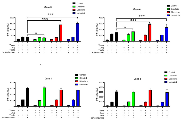
Figure 6.
In vitro treatment with anti-PD-1 with or without lenvatinib for CD8+ T cells of patients 1, 2, 4, and 5. All four patients were tested in vitro for 12 different conditions in triplicates. This figure shows the IFN-γ values in pg units upon 12 different conditions. mizoribine (red) and crizotinib (green) were used as positive and negative controls, respectively; the control (black) was untreated, and lenvatinib (blue) was the experimental test. A549 cells were first seeded on 96-well plates 24 h before adding the drugs. Then, drugs were added, and cells were incubated for 10 h. Media with or without anti-PD-1 antibody (pembrolizumab) containing the T cells of different patients was added after performing three PBSX1 washes for the whole 96-well plate. Human IFN-γ was performed using the collected media from the experimental wells and measured using the enzyme-linked immunosorbent assay (ELISA) method at a wavelength of 650 nm. A two-way analysis of variance (ANOVA) multiple comparisons test was performed among the groups for each patient individually, with three biologically independent counts in each group. *** p < 0.0001 for both the mizoribine- and the lenvatinib-treated groups in patients 4 and 5. All statistical data analysis was performed using GraphPad Prism, version 8.0.2.

