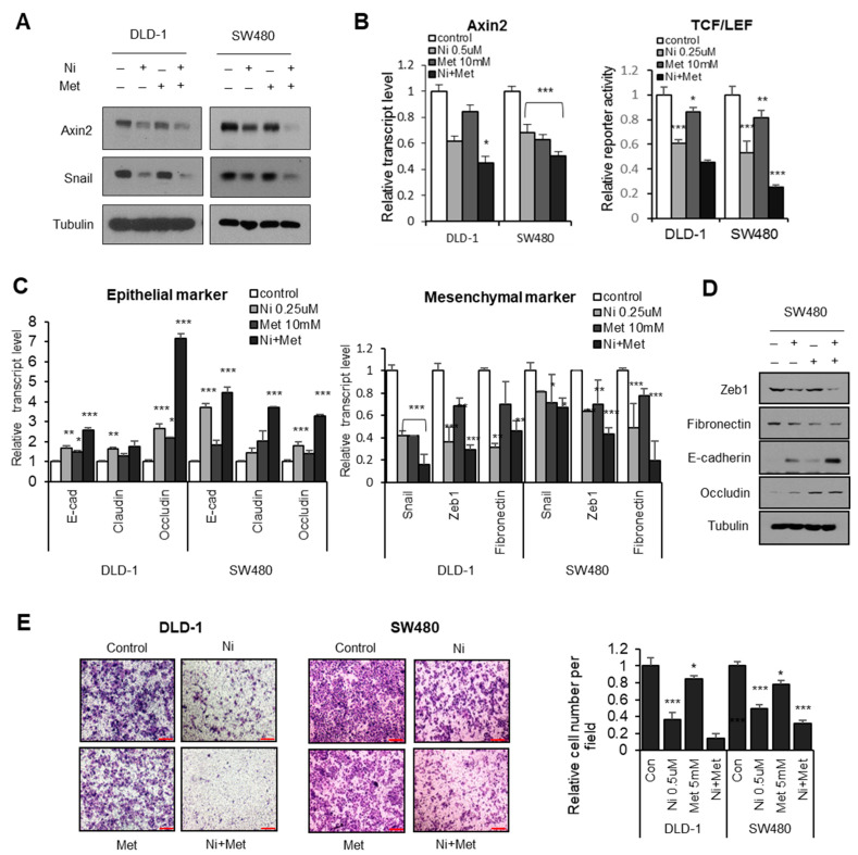
Figure 3.
Metformin potentiates niclosamide in canonical Wnt and Snail-mediated EMT. (A) The CRC cells were treated with either niclosamide (0.5 μM), metformin (5 mM), or a combination of the two for 16 h; endogenous Axin2 and Snail protein abundance were determined using immunoblot analysis. (B) Relative Axin2 transcript abundance (left) and TCF/LEF reporter (TOP flash) activity (right) in CRC cells. Statistical significances compared to control are denoted as * p < 0.05; ** p < 0.01; *** p < 0.001 by a two-tailed Student’s t-test. (C) Relative transcript abundance of epithelial markers (left) and mesenchymal genes (right) were determined by qRT-PCR in CRC cells treated with either niclosamide (0.25 μM), metformin (10 mM), or a combination of the two for 16 h. Statistical significances compared to control are denoted as * p < 0.05; ** p < 0.01; *** p < 0.001 by a two-tailed Student’s t-test. (D) The protein levels of epithelial and mesenechymal markers were determined in SW480 cells. (E) The migration ability of colon cancer cells treated with niclosamide (0.5 μM), metformin (10 mM), or a combination of the two was determined by transwell migration assay; scale bar, 200 μm. Statistical significances compared to control are denoted as * p < 0.05; *** p < 0.001 by a two-tailed Student’s t-test.

