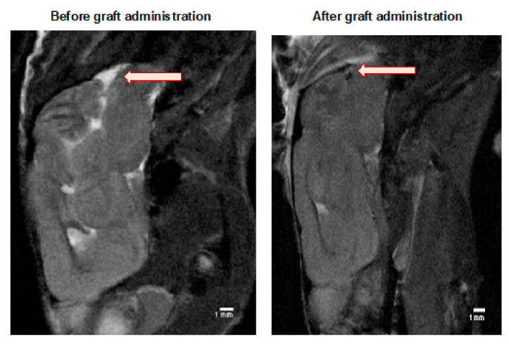
Figure 3.
MRI evaluation of the GRPs’ post-transplant localization in CNS: The MRI scan image of the representative (SCID) mouse at the day before (left picture) and a day after (right picture) of murine GRP transplantation into cisterna magna. The black dot (arrow), visible on the right picture shows the localization of SPIO-labelled cells. Abbreviations: GRP-glial-restricted progenitor cell; MRI-magnetic resonance imaging; SPIO-superparamagnetic iron oxide.

