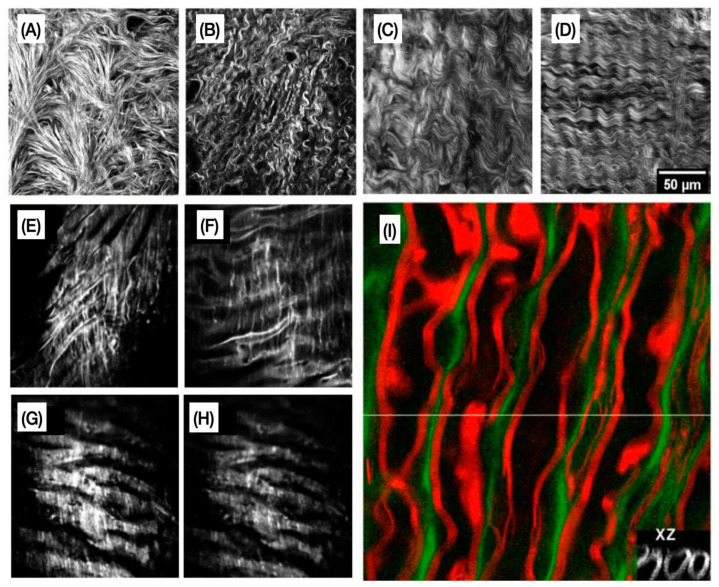
Figure 4.
Representative multiphoton microscopy images. (A–D) Second-harmonic generation (SHG) optical sections of collagen from the four categories of ovarian tissues [55]. (E–H) Examples of two-photon emission fluorescence (TPEF) images of elastic fibers acquired from the regions along the aorta of myocardial infarction-prone Watanabe heritable hyperlipidemic rabbits [56]. (I) Simultaneous coherent anti-Stokes Raman scattering (CARS) imaging of axonal myelin and TPEF imaging of Oregon green 488 is represented by red and green colors. The grayscale inset image is an XZ image showing the cross-section of axons [57]. Images reproduced with permission from [55,56,57].

