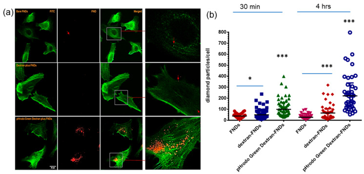
Figure 4.
Cellular uptake of diamond particles. (a) Confocal images of HeLa cells uptake FNDs, cells incubated with diamond particles for 4 h. green: pHrodo Green Dextran, DIC: Differential interference contrast, red: FNDs, the arrow indicates a diamond position diamond signal, scale bar: 20 µm. Arrows and zoomed in images used to indicate diamond location. (b) Analysis of diamond numbers in cells incubated with diamond particles for 30 min and 4 h. Three independent experiments were performed, 100 cells were counted for each group: bare FNDs, dextran plus FNDs, dextran-blocked cells first, pHrodo Green Dextran plus FNDs. * p < 0.05, *** p < 0.0001 as significant differences.

