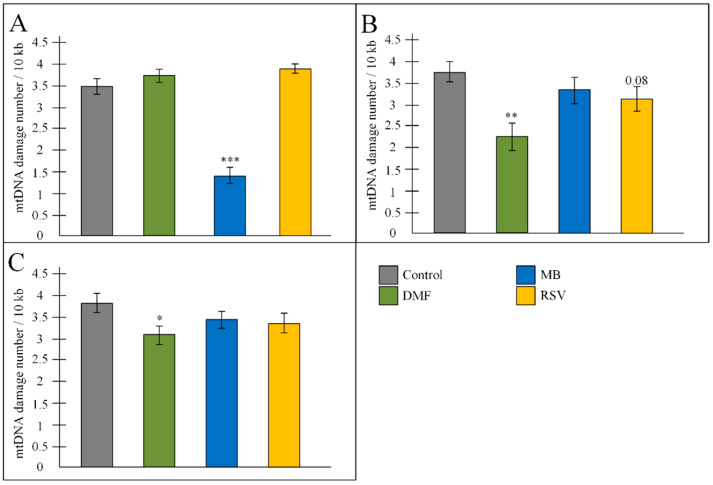
Figure 7.
Amount of mtDNA damage in the (A) hippocampus, (B) cortex, (C) mid-brain. The results expressed as means ± SEM. Control n = 8, DMF = 9, MB = 6, RSV = 7. The results expressed as means ± SEM. Control n = 8, DMF = 9, MB = 6, RSV = 8. * p < 0.05, ** p < 0.01, *** p < 0.001, comparison of the control group and treated groups using Tukey’s post-hoc test.

