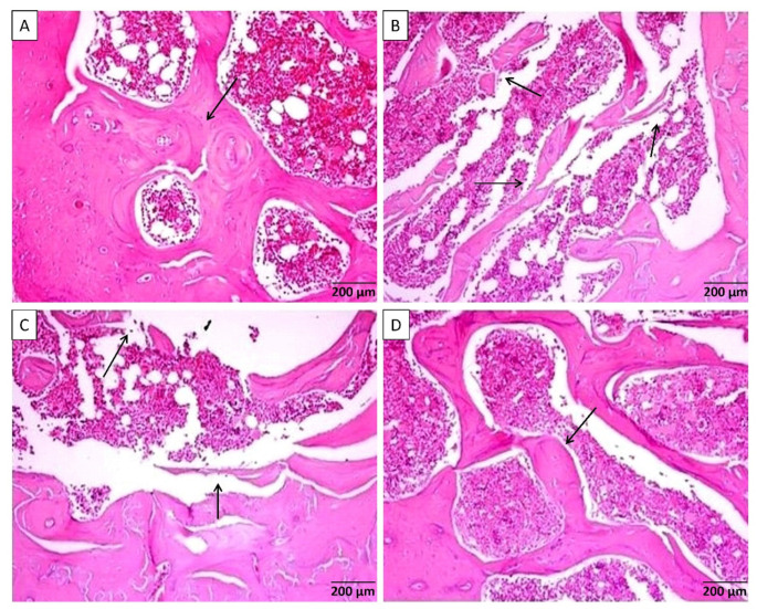
Figure 1.
(A) Rat femur sections from control group-I showed benign looking bone cortex and trabeculae [arrow] with normal mineralization separated by bone marrow element. (B) Sections from diabetic group-II with osteoporotic changes showing thickening of bone cortex associated with separated bone trabeculae [arrows] containing bone marrow element. (C) Rat femurs from group-III revealed fewer thinning of bone trabeculae [arrows] and some degree of separation seen containing bone marrow elements. (D) Microscopical description of group-IV femurs revealed well-formed bone trabeculae [arrow] with benign looking thickness and mineralization with joined trabeculae separated by bone marrow element.

