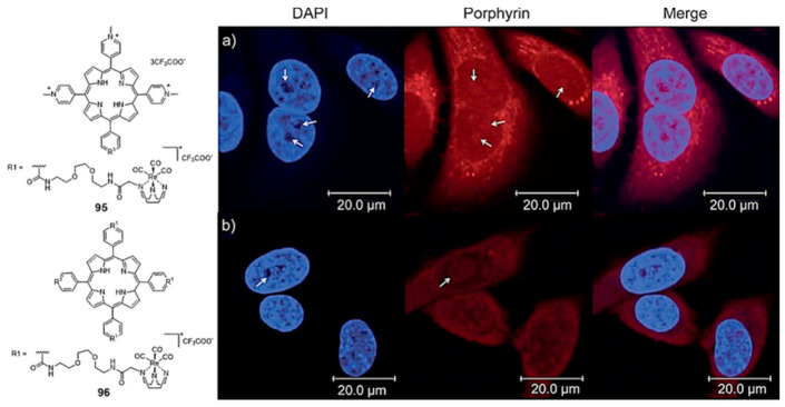
Figure 31.
Fluorescence confocal microscopy images of HeLa cells incubated for 2 h with tricarbonyl Re(I) complexes (95 (a) and 96 (b)) attached to a porphyrin scaffold and stained with DAPI. White arrows indicate the nucleoli. Reproduced with permission from ref. [151], published by John Wiley and Sons.

