
Figure 37.
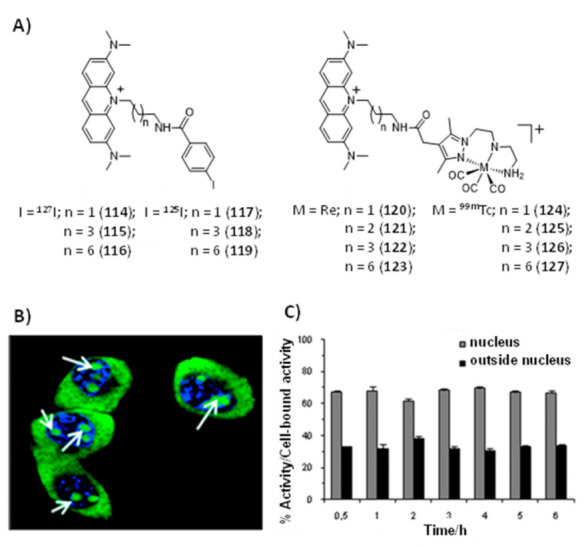
(A) Chemical structures of acridine orange derivatives containing natural elements (127I or Re) (114–116; 120–123) and Auger-emitting radionuclides (125I or 99mTc) (117–119; 124–127); (B) Uptake of the Re complex 121 in B16F1 murine melanoma cells, as evaluated by confocal fluorescence microscopy (blue: nuclei; white arrows: nucleoli); (C) Nuclear internalization of the 99mTc complex 125 in B16F1 murine melanoma cells, the activity values inside the nucleus and outside the nucleus are expressed as a percentage of cell-bound activity (mean ± standard deviation) [175,176].