Figure 3.
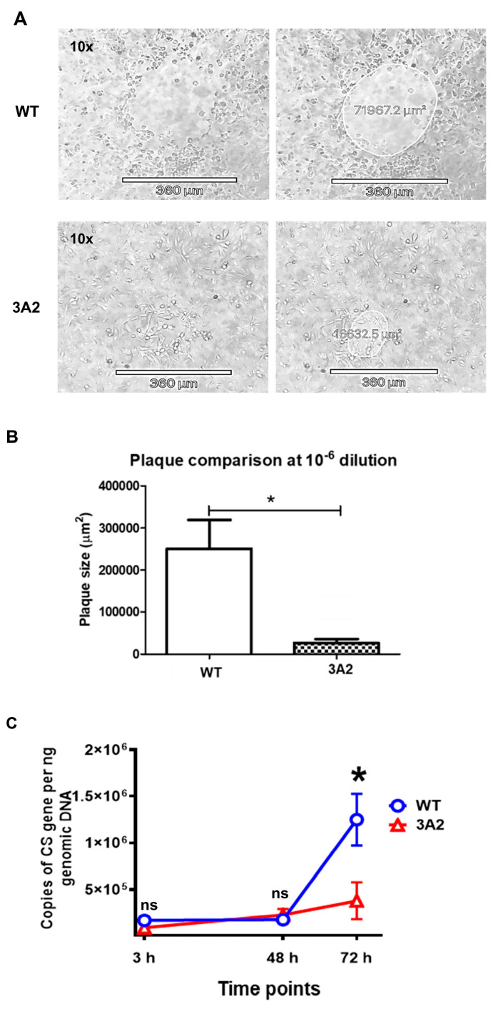
Evaluation of the virulence of R. parkeri 3A2 in vitro. Plaque assay was performed by the procedures described in Materials and Methods. The plaques generated by R. parkeri WT and 3A2 were monitored, imaged and measured. During days 3–5 p.i., the representative plaques generated by R. parkeri WT and 3A2 at the same dilution on the same day of infection are shown in (A). On the next day, plaques were determined and analyzed (B). (C) Human THP-1 cells were differentiated to macrophages using phorbol myristate acetate (PMA), and infected with R. parkeri WT and 3A2 at an MOI of 5. At 3 h, 48 h and 72 h p.i., the intracellular concentrations of R. parkeri WT and 3A2 were determined by quantitative real-time PCR amplifying the citrate synthase (CS) gene. Data represent two independent experiments with similar results. ns, not statistically significant. *, p < 0.05.

