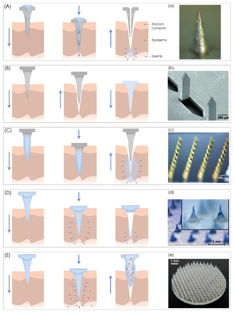
Figure 1.
A schematic representation of five different microneedle (MN) types for transdermal drug delivery. (A) Hollow MNs puncture the skin and release a liquid drug formulation through the needle lumen. (B) Solid MNs create microchannels in the skin and increase drug permeability. (C) Coated MNs enable drug dissolution into the skin from the coating film. (D) Dissolving MNs release the drug incorporated within the MNs. (E) Hydrogel MNs collect interstitial fluids and induce drug release through the swollen microprojections. (a) Bright-field microscopy (SZX 16, Olympus, Center Valley, PA, USA) image of hollow MNs. Reproduced with the permission from [80], Springer Nature, 2013. (b) SEM microscopy of solid MNs. Reproduced with the permission from [81], Elsevier, Amsterdam, The Netherlands, 2011. (c) bright-field microscopy (Olympus SZX12 stereo microscope, Olympus America) image of coated MNs. Reproduced with the permission from [37], Springer Nature, 2007. (d) microscope (STC-GE33A, SENTECH, Yokohama, Japan) image of dissolving MNs. Reproduced with the permission from [82], Elsevier, 2013. (e) picture of a hydrogel microneedle patch. Reproduced with the permission from [83], John Wiley & Sons—Books, 2015. The image was created with Adobe Illustrator CC (Version 23.0.1.; Adobe Inc., San Jose, CA, USA).

