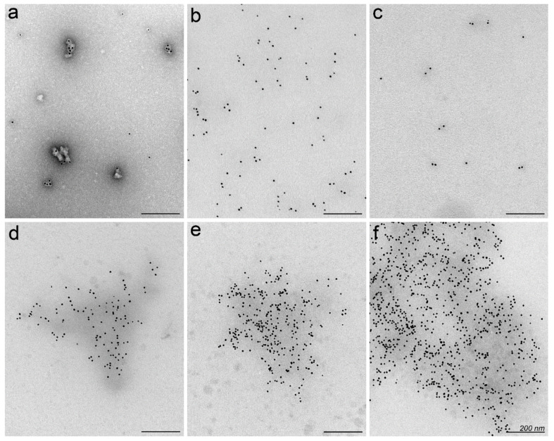
Figure 2.
Visualisation of PF14-SCO nanoparticles and protein corona by transmission electron microscopy (TEM). Complexes of PF14 and biotin-SCO labelled with colloidal gold of 10 nm diameter (black dots) were formed in water at PF14/SCO molar ratio 5. PF14-SCO complexes without protein corona negatively stained with uranyl acetate on TEM grid (a), and in unstained specimen (b). Biotin-SCO labelled with colloidal gold and stained with uranyl acetate (c). Protein corona forming in 50% bovine (d), murine (e) or human serum (f) clusters PF14-SCO nanoparticles to conglomerates with electron dense background. Scale bar: 200 nm. Representative images from three independent experiments are presented.

