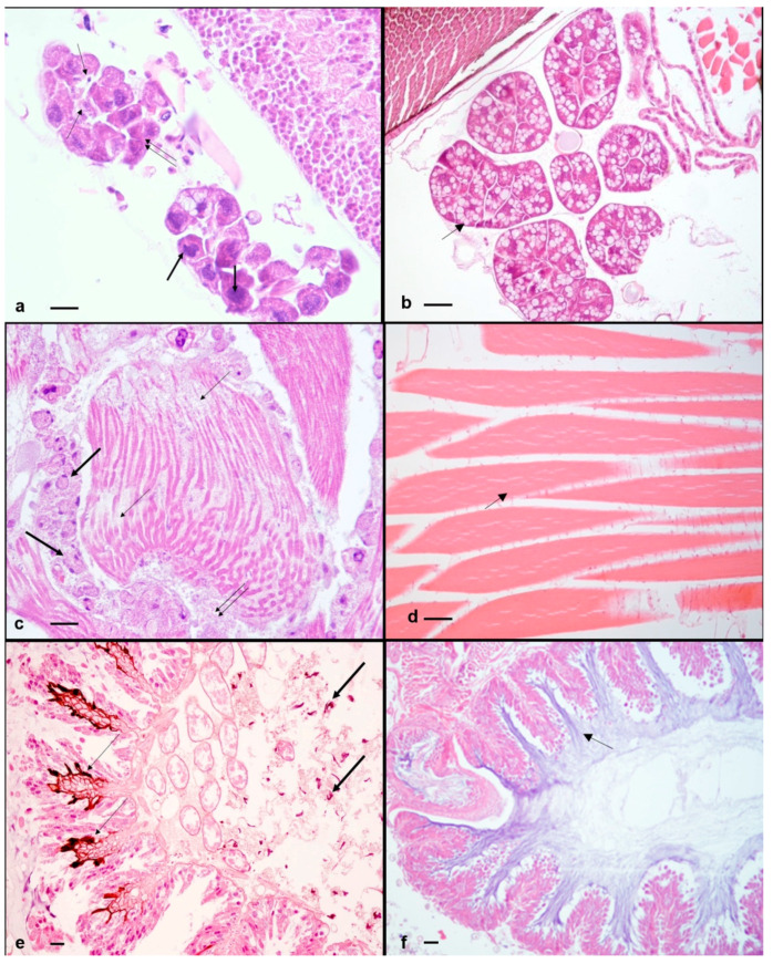
Figure 2.
A. mellifera with symptomatic and asymptomatic infection of DWV and confirmed presence of ABPV. (a) Symptomatic honeybee. Hypopharyngeal glands. Small irregular acini showing cells with hyperchromic nuclei (thick arrows), cytoplasm filled with few small vacuoles and eosinophilic granules (double arrow), plasmatocytes in the gland lumen (thin arrows). H-E. 400× (40× objective and 10× ocular). (b) Asymptomatic honeybee. Hypopharyngeal glands. Large acini showing cells with cytoplasm filled with numerous large vacuoles with clear foamy material (thin arrow). H-E. 400×. (40× objective and 10× ocular). (c) Symptomatic honeybee. Flight muscles. Fibers with few not completely formed myofibrils (thin arrows), numerous muscle-forming nuclei (thick arrows), trophocytes with nuclear fragmentation and eosinophilic material between the muscle fibers (double arrow). H-E. 400× (40× objective and 10× ocular). (d) Asymptomatic honeybee. Flight muscles. Numerous fibers with many well-formed myofibrils (thin arrow). No trophocytes are present. H-E. 400× (40× objective and 10×ocular). (e) Asymptomatic honeybee. Midgut. Melanin accumulation between the fold of the villi (thin arrows) and in the hemocele (thick arrows). H-E. 200× (20× objective and 10× ocular). (f) Symptomatic honeybee. Midgut. Absence of melanization and hemocytes. The midgut epithelium appears intact and the peritrophic membrane appears well lined (thin arrow). H-E. 200× (20× objective and 10× ocular). Scale bar: 50 μm.

