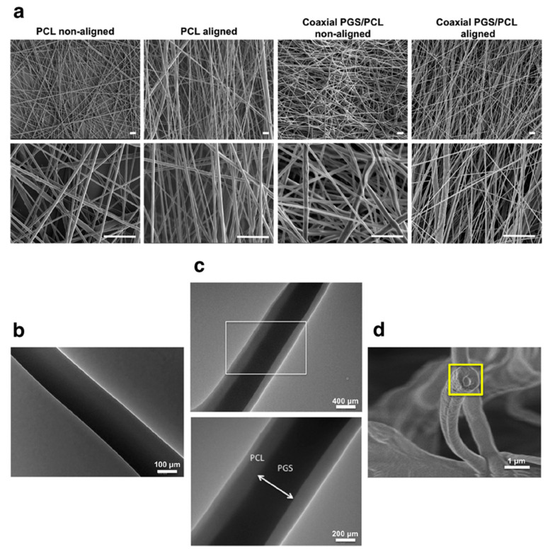
Figure 9.
Monolithic and core-shell electrospun fibres. Scanning electron microscopy (SEM) images (a) of aligned and non-aligned monolithic PCL and core-shell PGS-PCL fibres at two different magnifications. Scale bars: 5 μm. Transmission electron microscopy (TEM) images of (b) PCL monolithic fibres and (c) PGS-PCL core-shell fibres. The bottom panel in c is a magnification of the area within the white box in the top panel. The core-shell structure of PGS-PCL fibres was also confirmed by SEM imaging of the fibre cross-section (d, yellow box). Abbreviations: PCL—poly(ε-caprolactone); PGS—poly(glycerol sebacate). Adapted from [289] with permission from Elsevier. Copyright © 2019, Elsevier B.V.

