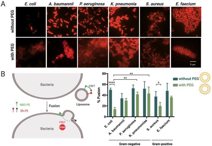
Figure 3.
Interaction and fusion of liposomes, with and without DSPE-PEG, with clinically relevant bacteria. (A) Representative epifluorescence microscopy pictures of bacteria incubated with Rh-labeled liposomes. Scale bar represents 5 μm. (B) Percentage of Rh/NBD liposomes fused with bacteria. Three independent experiments were done. Results are represented as mean values and respective standard deviations. Statistical differences are indicated when appropriate (p ≤ 0.0001, ****; p ≤ 0.01, **; and p ≤ 0.05, *). Figure 3B (left) was created with BioRender.com. (Accessed on 1 June 2021).

