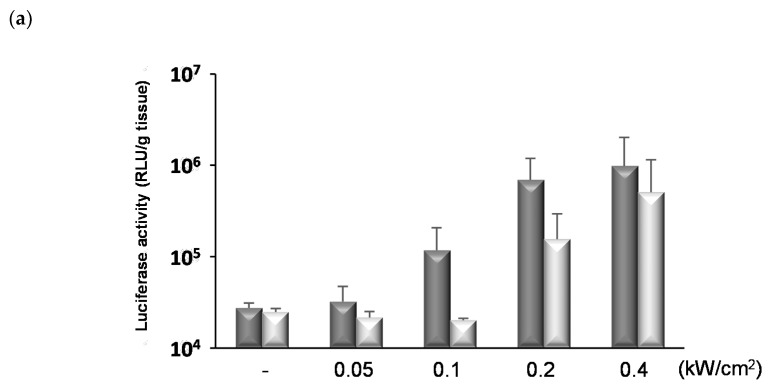
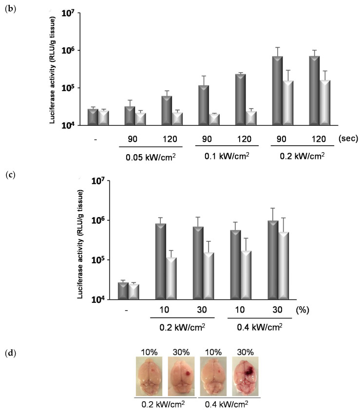
Figure 5.
The effects of FUS conditions on the gene transfection effects into the brain tissue. The ternary complexes (pDNA 48 μg/mouse, N/P 10, PEG-PEI:RVG-R9 = 8:2) and NBs (100 μg/mouse) were injected into the tail vein of mice and the right hemisphere was concurrently exposed to FUS. The FUS conditions for each study are as follows: (a) intensity: 0.05–0.4 kW/cm2, duty: 30% (sec), time: 90 s, (b) intensity: 0.05–0.2 kW/cm2, duty: 30% (sec), time: 90 or 120 s, (c,d) intensity: 0.2 or 0.4 kW/cm2, duty: 10 or 30% (sec), time: 90 s. One day after the transfection, the brain tissues were collected and the luciferase activity was analyzed. The bars show the mean and S.D. (n = 3–6). Dark gray column: the right hemisphere exposed to FUS. Light gray column: the left hemisphere unexposed to FUS. (d) The tissue damages by NBs and FUS 1 day after the transfection.


