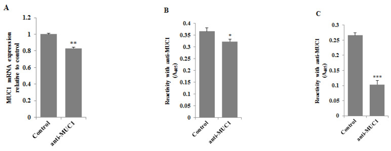
Figure 3.
The effect of anti-MUC1 mAb on MUC1 mRNA (A), MUC1 glycoprotein expression in cell lysates (B), and culture medium (C). The AGS gastric cancer cells were incubated for 24 h with anti-MUC1 (5 μg/mL). mRNA was assessed by RT-PCR. The result is shown as a relative fold change in mRNA expression of the gene in comparison to the gene in controls where expression was set at 1 ± SD are the mean of triplicate cultures. ** p < 0.01. MUC1 glycoprotein expression was determined by ELISA tests. The results are expressed as absorbance at 405 nm after reactivity with anti-MUC1 monoclonal antibody (BC2 clone). Values ± SD are the mean from three independent assays. * p < 0.05; *** p < 0.001.

