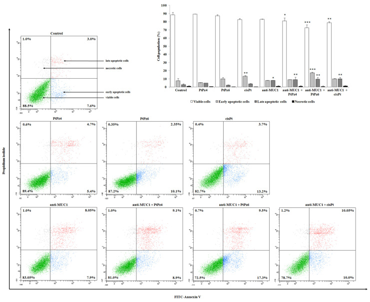
Figure 5.
Flow cytometry analysis of AGS gastric cancer cells after 24 h incubation with PtPz4 (10 μM), PtPz6 (10 μM), cisPt (10 μM), anti-MUC1 (5 μg/mL), PtPz4 + anti-MUC1 (10 μM + 5 μg/mL), PtPz6 + anti-MUC1 (10 μM + 5 μg/mL), and cisPt + anti-MUC1 (10 μM + 5 μg/mL) and successive staining with Annexin V and propidium iodide. The data are presented as mean percentage values from three independent experiments (n = 3) performed in duplicate. * p < 0.05; ** p < 0.01; *** p < 0.001 versus control group.

