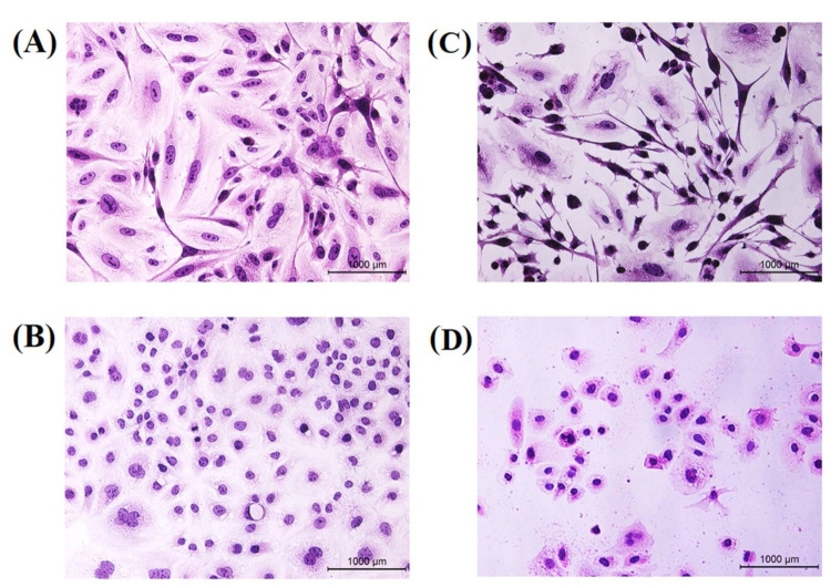
Figure 12.
Morphological changes of hematoxylin and eosin. MCF-7 cell line (upper lane) and CAL-51 cell line (lower lane) were treated with IC50 concentration of TAM/RES–LbL-LCNPs for 24 h. (A,B) Non-treated cells show normal structure without prominent apoptosis at MCF-7. CAL-51, respectively (C,D) Cells treated with TAM/RES–LbL-LCNPs; apoptotic cells show condensed, and fragmented nuclei were recorded at MCF-7. CAL-51, respectively.

