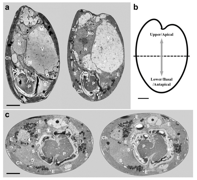
Figure 5.
Micrographs of Tetraselmis jejuensis obtained using transmission electron microscopy. (a) Longitudinal sections showing several organelles inside the protoplasm, including chloroplasts (Ch), eyespot granules (E), mitochondria (M), nucleus (N), nucleolus (No), pyrenoid (P), starch (S), and vacuoles (V). The pyrenoid was invaded by both cytoplasmic channels comprising electron-dense material separated from the cytoplasm and canaliculi traversing it in opposite directions (black arrow). (b) Schematic drawing of longitudinal section indicating the upper/apical and lower/basal/antapical positions. (c) Transverse sections showing the position and structure of chloroplast (Ch), eyespot granules (E), pyrenoid (P), and starch (S). Scale bars = 2 µm.

