Figure 1.
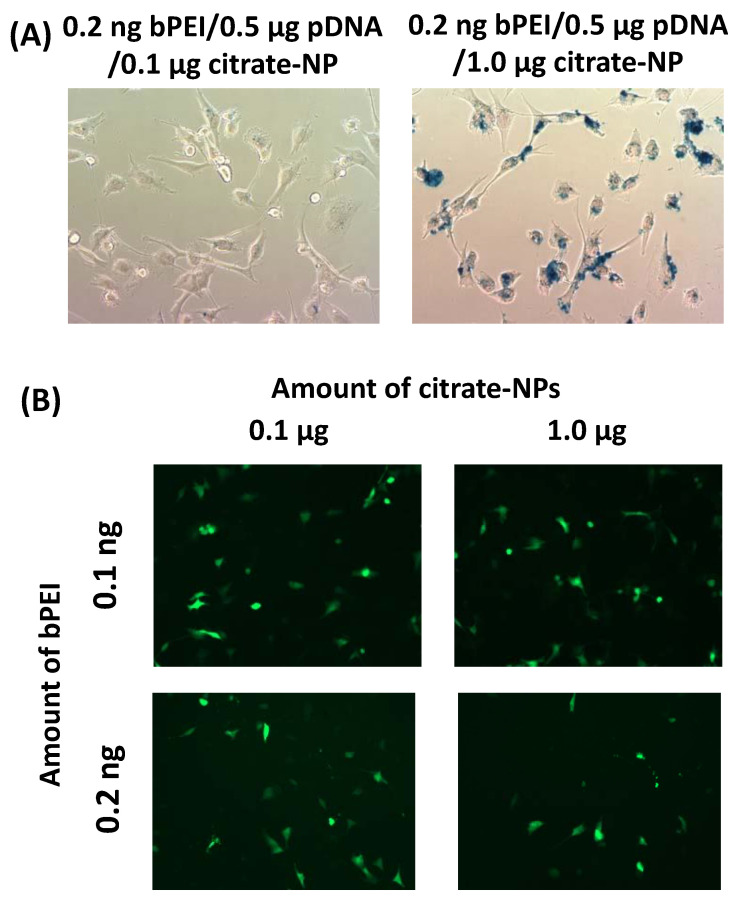
(A) Prussian staining images of two different composites 0.2 ng branched bPEI/0.5 μg pEGFP-C1 pDNA/0.1 μg citrate-NP and 0.2 ng branched bPEI/0.5 μg pEGFP-C1 pDNA/1.0 μg citrate-NP 24 h after incubation with each composite for 5 h in U87MG cells. (B) GFP fluorescence images of four different composites with a fixed amount of 0.5 μg pEGFP-C1 pDNA 24 h after incubation with each composite for 5 h in U87MG cells.

