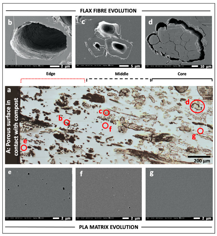
Figure 3.
(a) Optical micrograph of the investigated face C (see Figure 1), which is divided into edge, middle and core areas following visual observation of fibre degradation; (b–d) SEM images of fibres collected in edge, middle and core, respectively. The complete degradation of the fibres leaves phantom cavities with the same shape of the degraded fibre (b). Different stages of degradation can be recognised in (c). In (d), fibres in core appear intact; (e–f) SEM images of PLA matrix investigated in edge, middle and core, respectively. Some porosities visible in edge (e) and middle (f), probably due to degradation, are absent in core (g). For each SEM image, the corresponding investigation area is indicated with red circles in (a).

