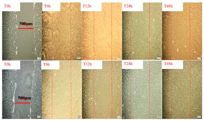
Figure 5.
Migration of dermal fibroblasts after treatment with mixed extract of Trifolium pretense and Ocimum basilicum (lower line) compared to the control (upper line) monitored after different times intervals under light microscopy (objective 20×). Scale bar: 100 μm. The edge of initial pseudo-wound area is labeled in red, in order to emphasize the progressive covering of the area, during 48 h incubation (unpublished results).

