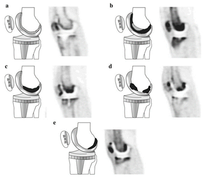
Figure 6.
Diagrams and representative PET images showing the five types of 18F-NaF uptake pattern in the femoral component of knee prostheses. Black areas represent areas of severely increased uptake and shaded areas represent slightly increased uptake of radiotracer. Adapted with permission from Son et al. (2016) [50] Copyright Springer Nature, 2016.

