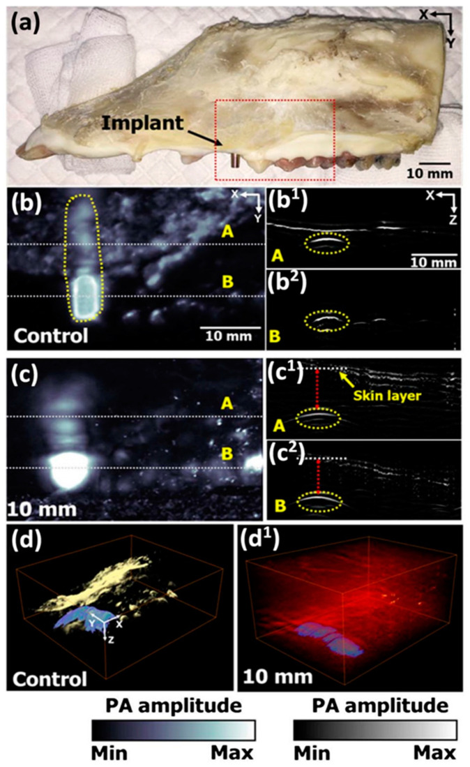
Figure 7.
Ex vivo PA image of porcine jawbone with titanium implant abutment (EZ Post, MIEP3525HT, Megagen, Korea) and a fixture (MIIF3008C, Megagen, Korea). (a) Jawbone specimen. (b) PA image at 1064 nm excitation. (c) PA MAP images from (b) under 10 mm of chicken tissue. (d) 3D render of bare bone specimen (d) and under 10 mm chicken tissue (d1). (b1,b2) and (c1,c2) are cross-sectional PA images of the dashed line areas in corresponding (b,c) images.Reprinted with permission form Lee et al. (2017) [59] Copyright The Optical Society of America, 2017.

