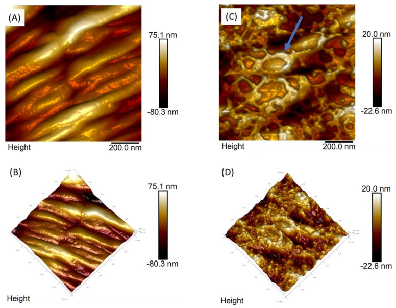
Figure 4.
Two-dimensional (2D) and 3D analyses of the L2 cuticle of H. contortus by AFM. Non-treated H. contortus AFM height image in L2 stage (A) and its 3D visualization (B), H. contortus L2 treated with RcAlb-PepIII (C) and its three-dimensional visualization (D). In the images (C,D), it is possible to observe the alteration promoted by the attachment of the peptide solution to the cuticle in the L2 nematode. The blue arrow points to the presence of structures on the nematode surface observed after their exposure to peptide solution. Both images have a 1 micrometer square scan.

