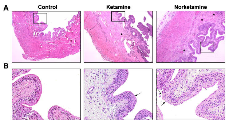
Figure 3.
Haematoxylin and eosin staining of the bladder from each group. The images of the upper row (A) were taken at 40× magnification and images of the lower row (B) were taken at 200× magnification. The star marks indicate the edema sites in the lamina propria, and the damaged glycosaminoglycan (GAG) layer shows in the edge of the epithelial layer (arrows).

