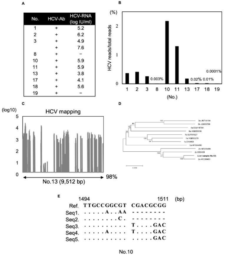
Figure 4.
Detection of HCV reads obtained from recipient liver samples. (A) Recipient liver sample list of HCV infection previously with HCV-Ab positive, HCV-RNA was evaluated by preoperative blood test. (B) HCV reads compared to total reads obtained from each liver sample were shown in bar graph. (C) HCV reads from a liver sample (No.13) were mapped. A bar graph is shown and the percentage of the total number of reads that were mapped to the HCV genome is indicated. (D) Phylogenetic tree analysis in the NS5B region by the viral sequence obtained from HCV reads (HCV No. 13). (E) Analysis of quasi-species using HCV reads obtained from liver samples (No.10). Alignment of HVR1 (1494–1511 bp). Nucleotide positions are numbered according to GenBank accession number NC004102. The five major sequences are listed.

