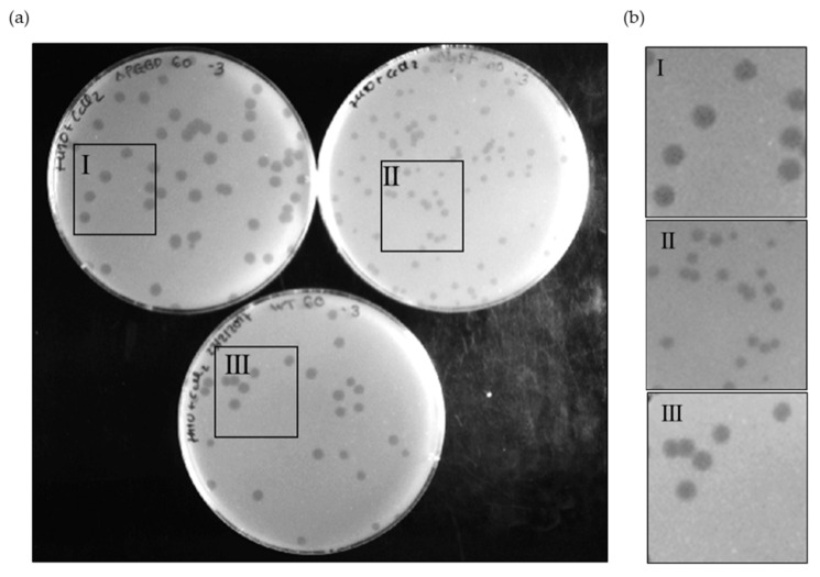
Figure 4.
Phage plaque morphology of Ms6lysBΔPGBD (I), Ms6ΔlysB (II) and Ms6wt (III) on a lawn of M. smegmatis (a), and 2× zoom of each highlighted area (b). The mutant Ms6lysBΔPGBD forms slightly larger plaques than the Ms6wt, while Ms6ΔlysB, in turn, forms the smallest plaques. The pictures were taken at 24 h post infection.

