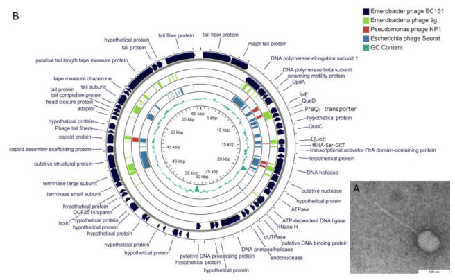
Figure 1.
Phage EC151 characteristics. (A) Electron micrograph of phage EC151 particle. Transmission electron microscopy, negative staining with 1% uranyl acetate; (B) genome map of Enterobacter phage EC151 constructed using the CGView server. For sequence similarity comparison, TBLASTX was used versus Escherichia phage 9 g (light green), Pseudomonas phage NP1 (brown), and Escherichia phage Seurat (cyan).

