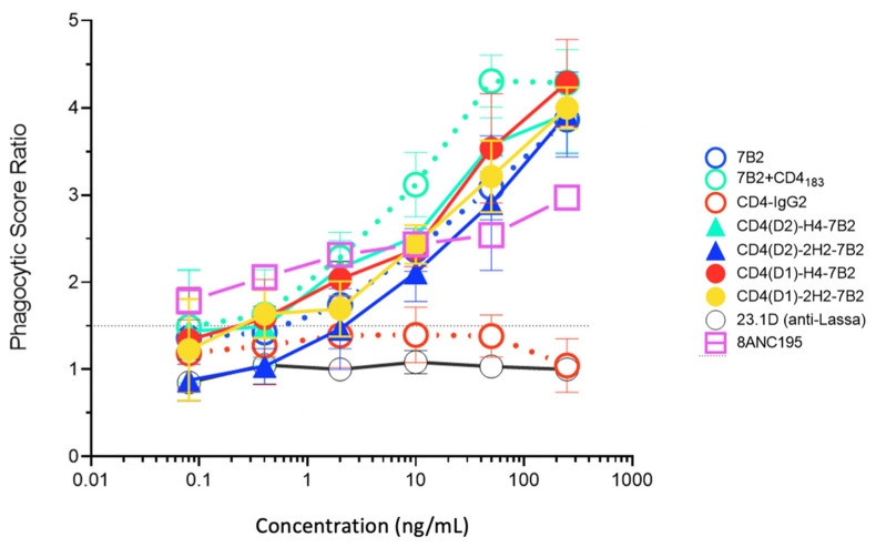
Figure 6.
Antibody-dependent phagocytosis with panel mAbs and Ig-adhesins. We quantified the ingestion of gp140-coated fluorescent beads by THP-1 cells. Beads were opsonized with the indicated Ab. The novel Ig-adhesins are represented as solid lines and symbols, while the controls are dotted lines and open symbols. For each concentration, the phagocytic score and SEM are shown. When no error bars are visible, they are smaller than the symbol. The horizontal line indicates the cutoff for significance. The data shown are representative of three separate experiments.

