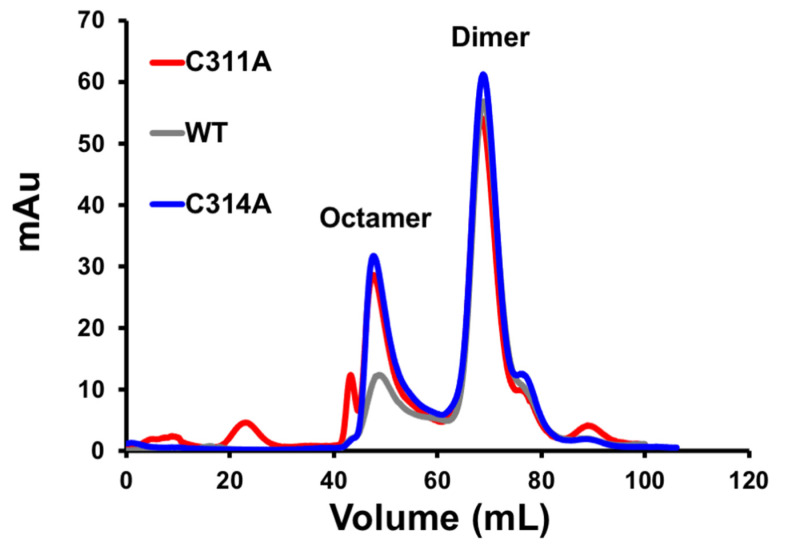
Figure 6.
Dimer and octamer assessment of VP40 and single cysteine mutations in vitro. Size exclusion chromatogram trace of WT, C311A, and C314A purified VP40 protein shows similar formation of the dimer in solution compared to WT (only dimers used for lipid-binding assays). C311A and C314A both displayed an increased propensity towards the octamer form compared to WT VP40. The double mutant, C311A/C314A is not shown but demonstrated similar dimer and octamer peaks analogous to the single mutants.

