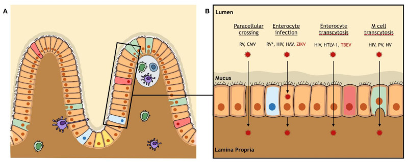
Figure 5.
The intestinal barrier: anatomy and viral crossing. (A) The intestinal barrier is a monostratified epithelium composed of different cell types that all differentiate from pluripotent stem cells (yellow) located at the bottom of the crypts: enterocytes (orange), goblet cells (pink), Paneth cells (blue), and microfold cells (M cells) (green). Underlying the epithelium, the gut-associated lymphoid tissue is composed of free and anchored immune cells. Over the lymphoid follicles, the follicle-associated epithelium is enriched by M cells. (B) The intestinal barrier can be crossed by paracellular crossing by Rotavirus (RV), by enterocyte infection by RV (*: after paracellular crossing), Human immunodeficiency virus (HIV), Hepatitis A virus (HAV), and Zika virus (ZIKV), by transcytosis through enterocytes by HIV, type 1 Human T-cell lymphotropic virus (HTLV-1), and Tick-borne encephalitis virus (TBEV), or by transcytosis through M cells by HIV, Poliovirus (PV), and Norovirus (NV). The only arboviruses (red) that were reported to cross the intestinal barrier are TBEV and ZIKV.

