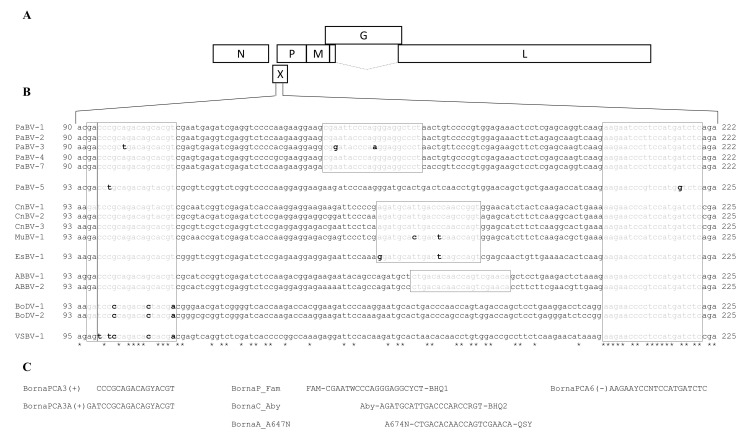
Figure 2.
Primer and probe design. (A). Schematic presentation of the orthobornavirus genome. (B). Alignment of the partial orthobornavirus X/P gene sequences (positions 90/93 to 222/225). Boxes: sequences corresponding to the primer and probe binding regions; grey: nucleotides matching the primer/probe sequence; black, boldface: nucleotides mismatching with the primer/probe sequence. PaBV-6 and PaBV-8 are not included in this alignment, since no X gene sequences are available for these viruses. (C). Primer and probe sequences.

