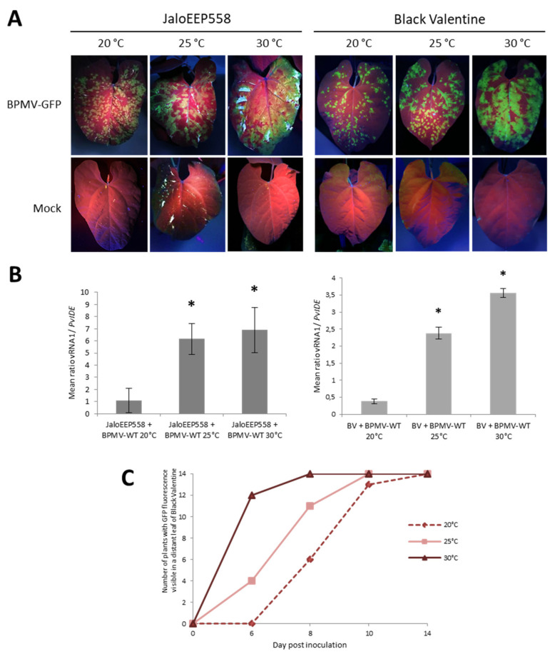
Figure 4.
Elevated temperatures promote BPMV infection in the inoculated leaf and BPMV systemic spreading in whole plants of two susceptible genotypes of P. vulgaris: (A) Representative pictures of four inoculated leaves from four different plants of either JaloEEP558 or Black Valentine (left and right panel, respectively) at 20 °C, 25 °C and 30 °C, 7 days post-inoculation (dpi) with BPMV-GFP (upper panel) and Mock (lower panel). BPMV-GFP was detected under UV light. This experiment was performed at least three times with similar results. (B) Quantification of BPMV titer at 7 dpi in four BPMV-WT-inoculated leaves of four different plants of JaloEEP558 or Black Valentine (BV) by calculation of the relative ratio of BPMV RNA1 to plant mRNA of PvIde using a quantitative RT-PCR procedure. Asteriks indicate significant differences between the tested temperature and 20 °C, the control temperature (t-test, p-value < 0,05). No significant difference of viral titer was found between the two elevated temperatures 25 °C and 30 °C. Data are mean ratios ± SD of four biological replicates. (C) BPMV-GFP systemic spreading in whole plants of Black Valentine at 20 °C, 25 °C, and 30 °C. The graph represents the number of plants with GFP fluorescence visible in a distant leaf scored at four dates after inoculation with BPMV-GFP: 0, 6, 8, 10, and 14 dpi on a total of fourteen plants per temperature assay. BPMV-GFP was detected under UV light. This experiment was performed twice with similar results.

