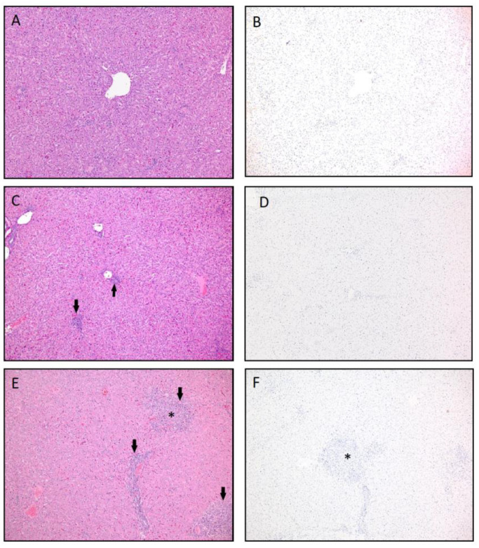
Figure 7.
Liver histopathology and viral antigen immunohistochemistry (IHC) representative of absent to minimal to moderate hepatic inflammation in vaccinated animals. (A) Hematoxylin and eosin stain (H&E) of normal liver. H score was 0 = no lesions (Group 3, Animal #3, 10 dpc). (B) No viral antigen was present by IHC (Group 3, Animal #3, 10 dpc). (C) H&E of the liver with low numbers of periportal lymphocytes and plasma cells (arrows). H score was 1 = minimal inflammation (Group 2, Animal #9, 10 dpc). (D) The tissue was also negative for viral antigen by IHC (Group 2, Animal #9, 10 dpc). (E) H&E of multifocal inflammation in portal tracts (arrow) as well as a focus of inflammation with hepatocyte loss (*). H score was 2.5 = moderate inflammation (Group 4, Animal #14, 7 dpc). (F) IHC was negative for viral antigen (Group 4, Animal #14, 7 dpc). 100× magnification. The H score is the hepatic histopathology lesion score based on the lesion character, amount of hepatic parenchyma affected and lesion severity on a scale from 0 (no lesions) to 4 (severe lesions_ (see Table 1)).

