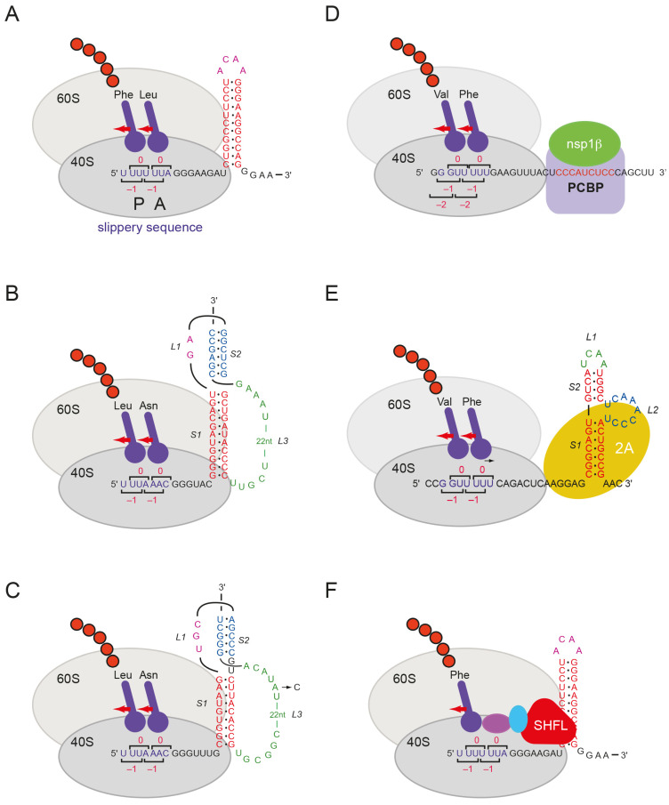
Figure 1.
Modulators of programmed –1 ribosomal frameshifting. Cartoon drawings of ribosomes encountering sites of PRF. Stimulators: (A) HIV stem-loop; (B) IBV pseudoknot; (C) SARS-CoV-2 pseudoknot (SARS-CoV sequence identical in the region shown except for an (A) to (C) transversion in loop 3 (L3) as indicated); (D) PRRSV complex of virus (nsp1β) and host protein (PCBP); (E) Cardiovirus (EMCV) stimulatory RNA complexed with viral 2A protein. Repressor: (F) SHFL shown bound at the HIV-1 signal. Purple and cyan ovals represent eRF1 and eRF3 (in complex).

